Early detection of bladder cancer
Photodynamic diagnostics (PDD) in flexible endoscopy technology, Munich
Introduction
As with any form of cancer, the earlier bladder cancer is detected, the better the chances of cure. We are the first and only practice in southern Germany to use the photodynamic diagnostic (PDD) method, which is otherwise only used in hospitals, for the early detection and clarification of bladder cancer in flexible endoscopy technology on an outpatient basis in our own practice.
The advantage of photodynamic diagnostics is that the cancer virtually exposes itself through a fluorescent blue light.
Urinary bladder cancer is the fifth most common cancer in men and the tenth most common cancer in women. In urology, it is among the third most common urological cancer entity (cancer type). The main risk factor for developing bladder cancer is smoking, although passive smoking also increases the risk of developing a bladder tumor. Urinary bladder cancer is considered the second most common cancer in smokers after bronchial cancer.
In its early stages, bladder cancer usually causes little or no symptoms. A first alarm sign can be the painless appearance of blood in the urine (painless hematuria), which can be detected microscopically (microhematuria) or can already be seen with the naked eye (macrohematuria).
Risks for the development of bladder cancer
In addition to smoking, workers in the dye industry, chemical and petrochemical industries, and tar-processing plants are at significantly higher risk of urinary bladder cancer due to high exposure to biogenic amines.
Excessive exposure to radiation after external radiation therapy in the pelvis as well as chronic urinary bladder infections, urinary bladder voiding disorders, cytostatics (chemotherapeutic drugs) can also cause bladder cancer.
In tropical and subtropical countries (for example in Egypt), urinary bladder carcinoma can occur as a late consequence of schistosomiasis (tropical infectious disease). This disease is caused by a freshwater-dwelling parasite (sucking worm) that enters the human organism through the skin, spreads as a host through the bloodstream, and lodges in the human urinary bladder wall. Within the urinary bladder wall, this parasite can cause bladder cancer.
Symptoms
In early stages, as previously stated, bladder cancer causes little or no discomfort. The painless appearance of blood in the urine (painless hematuria) can be a first alarm sign. This can be detected microscopically (microhematuria) or already with the naked eye (macrohematuria).
Likewise - then usually with pain during urination - blood in the urine can also be a sign of other, usually inflammatory diseases with a bacterial cause in the upper and/or lower urinary tract (kidneys/ureter/bladder/urethra).
Diagnostics with indication of bladder cancer
An essential part of the diagnosis of suspected bladder cancer is urinalysis, sonography (ultrasound examination), digital X-ray examination of the kidneys and ureters, and cystoscopy.
All these examinations, in particular the radiological imaging of the urinary tract, are carried out in our practice using the latest digital 3D X-ray technology.
Because bladder cancer often grows multilocular (i.e. at several sites within the bladder) and cannot always be detected by normal white-light endoscopy during initial diagnosis as well as during recurrence clarification and is thus overlooked, photodynamic diagnostics (PDD) offers a decisive advantage: cancer areas can be visualized in color and thus more easily distinguished from normal bladder mucosa.
![[Translate to Englisch:] pdd-diagnostik blasenkrebs muenchen](/typo3conf/ext/min_interchange/Resources/Public/Images/invis.gif)
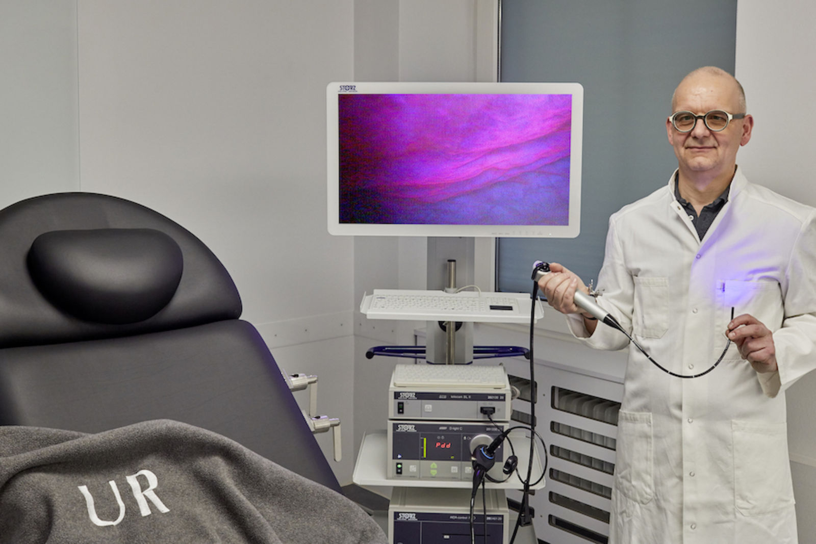
Photodynamic diagnostics (PDD)
Photodynamic diagnostics is a minimally invasive (without surgical intervention) diagnostic option for the detection of primarily inconspicuous (invisible) bladder cancer.
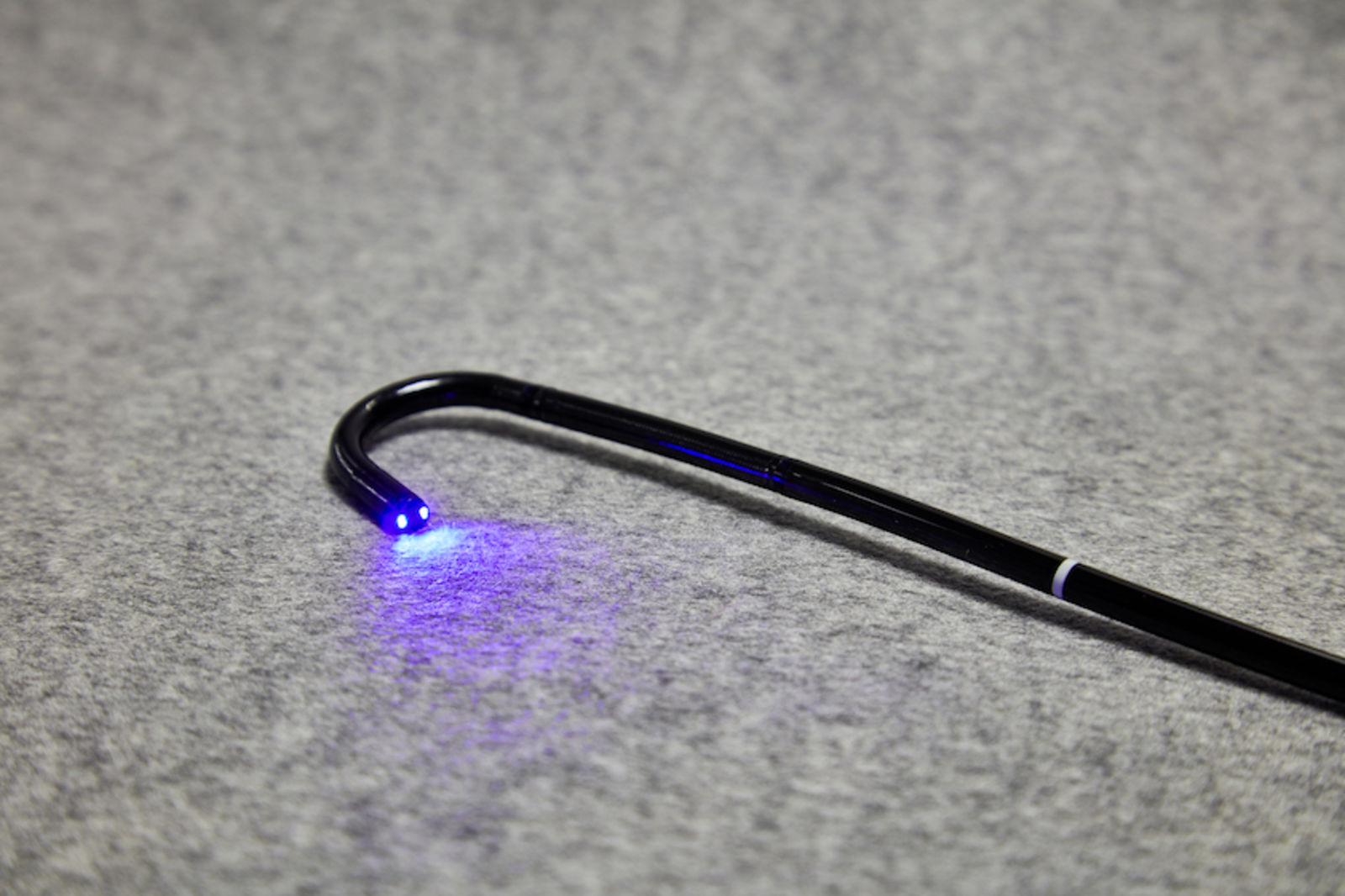
The diagnostic principle of this technique is based on the fact that cancer cells in the urinary bladder can be selectively stained with a substance that is introduced into the bladder via a thin catheter under local anesthesia approximately 60 minutes before the examination. During the subsequent examination, the urinary bladder is illuminated with ultraviolet light via an endoscope. Cancerous areas in the mucous membrane of the urinary bladder are highlighted in red and can be selectively removed with an electric snare or laser beam.
Tumor cells that are overlooked under conventional white light endoscopy can be detected much better with the PDD method.
The advantage of this technique is that the risk of overlooking tumors in the bladder can be significantly reduced. Particularly in recurrence diagnostics - i.e. in a control endoscopy after bladder tumor removal has already been performed - PDD offers the advantage that recurrent tumors can be detected better and thus earlier.
Due to the fact that bladder cancer can only be cured in its early stages by consistent and complete transurethral resection (removal of the tumor endoscopically using a resectoscope via the urethra), PDD offers the decisive advantage that the probability of missing tumor tissue is very low.
As part of his surgical activities, Dr. Friedemann Meisse is also able to remove bladder tumor tissue transurethrally under PDD control.
Advantages of flexible technology
While conventional "rigid" photodynamic diagnostics use unbendable metal optics, which usually require the patient to undergo a urinary bladder endoscopy under general anesthesia in order to avoid unnecessary examination pain, the flexible PDD technique with the aid of bendable and thus tissue-conserving endoscopes does not require general anesthesia and can therefore be performed on an outpatient basis under local anesthesia. This saves the patient hospital stays and thus time and also costs.
Special service: we offer three anesthetic options
In addition to local anesthesia, PDD can also be performed under general anesthesia and sedation (combination of sedation and local anesthesia).
We perform 98% of photodynamic diagnostics in the practice under local anesthesia. If you as a patient wish to have the examination performed under general anesthesia, we will perform the examination in the IATROS clinic. You will then be asleep during the examination, will be ventilated and will not notice anything.
In addition to local and general anesthesia, we offer a third version in our practice - endoscopy under sedoanalgesia. It is known to many from colonoscopy, where it is often used. In sedoanalgesia, the patient is asleep but not ventilated as in general anesthesia. This has the advantage that they are awake and mobile again more quickly after the procedure.
Lesen Sie auch: Früherkennung und umfassende Abklärung von Blasenkrebs mithilfe der flexiblen Photodynamischen Diagnostik (PDD)
Das besondere Verfahren der flexiblen Photodynamischen Endoskopiediagnostik (PDD) ermöglicht ein so frühzeitiges und differenziertes Erkennen von Harnblasentumoren wie es mit der herkömmlichen Methode der Weißlicht-Blasenspiegelung nicht möglich ist. Dr. Friedemann Meisse ist der erste niedergelassene Facharzt für Urologie in Süddeutschland, der diese Diagnostik in Form der flexiblen Photodynamsichen Diagnostik durchführt = Eine ambulante Durchführung OHNE Vollnarkose. Früherkennung - differenzierte Diagnostik bei Blasenkrebs - München